Medical Imaging comparison
페이지 정보
작성자 : Eden
조회수 : 8회
작성일 : 25-04-23 03:42
본문
When it comes to medical imaging technologies, several techniques are available to help treat and understand various health conditions. Two of the most prominent imaging modalities used today are nuclear heart scans and Magnetic Resonance Sequences, while both techniques are non-invasive and are used to create detailed images of the internal organs or structures, they differ in terms of their underlying principles, application areas, advantages, and limitations. In this article, we will discuss the comparisons between nuclear heart scans and MRI scans, helping you decide which technology is more suitable for your needs.
A nuclear heart scan, or myocardial perfusion scan, uses small amounts of radioactive compound or tracer to generate images of the heart. This imaging technology is commonly used to diagnose coronary artery disease, spot heart attack damage, and improve coronary artery bypass grafting. During a nuclear heart scan, a small amount of radioactive material is injected into the bloodstream. As the tracer travels through the body, it collects in the heart tissue and releases radiation. Specialized cameras then detect this emitted radiation, creating detailed images of blood flow and heart operation.
On the other hand, an MRI scan uses radio waves and a strong to produce images of the body's internal structures. The procedure involves lying on a table that slides into the MRI machine, which then uses a powerful magnetic field and radio waves to generate signals. These signals are picked up by sensors and combined with additional data from gradations in magnetic fields or imaging techniques like dynamic susceptibility contrast, that reveal detailed insights about the body's internal structures.
One of the main differences between nuclear heart scans and MRI scans is the type of results they produce. Nuclear heart scans mainly concentrate on blood flow and cardiac function, making them more suitable for diagnosing cardiovascular conditions. MRI scans, on the other hand, can capture detailed images of soft tissues, including the brain, liver, kidneys, and other organs. This allows MRI scans to be used for a wider range of applications, including identifying musculoskeletal injuries, tumors, and other conditions.
Another key difference between the two imaging technologies is the level of ionizing radiation they require. Nuclear heart scans need the use of radioactive material, which carries a small risk of radiation exposure. MRI scans, on the other hand, are free from ionizing radiation, making them a safer choice for expectant mothers and patients who require recurring imaging procedures.
When it comes to pre-imaging preparation, اسکن قلب nuclear heart scans require patients to abstain from food for a few hours, often restrict caffeine intake, and may require additional preparation. MRI scans have fewer restrictions, but individuals with metal implants, such as pacemakers, or certain medical diseases, such as claustrophobia, may need to complete additional testing or consult with a doctor before undergoing the procedure.
In conclusion, both nuclear heart scans and MRI scans are advantageous medical imaging technologies that provide valuable diagnostic results. While nuclear heart scans offer detailed images of cardiac condition and blood flow, MRI scans can obtain soft tissue details, including brain, liver, and other structures, making them more versatile and widely practical. Understanding the differences and similarities between these imaging technologies can help you select the right diagnostic tool for your particular medical needs.
When discussing with your doctor regarding nuclear imaging versus MRI, a conversation centered on the specifics of the imaging is key. Keeping these differences in mind will allow you to inquire your physician inquiries and ultimately reach a decision to the proper course of action.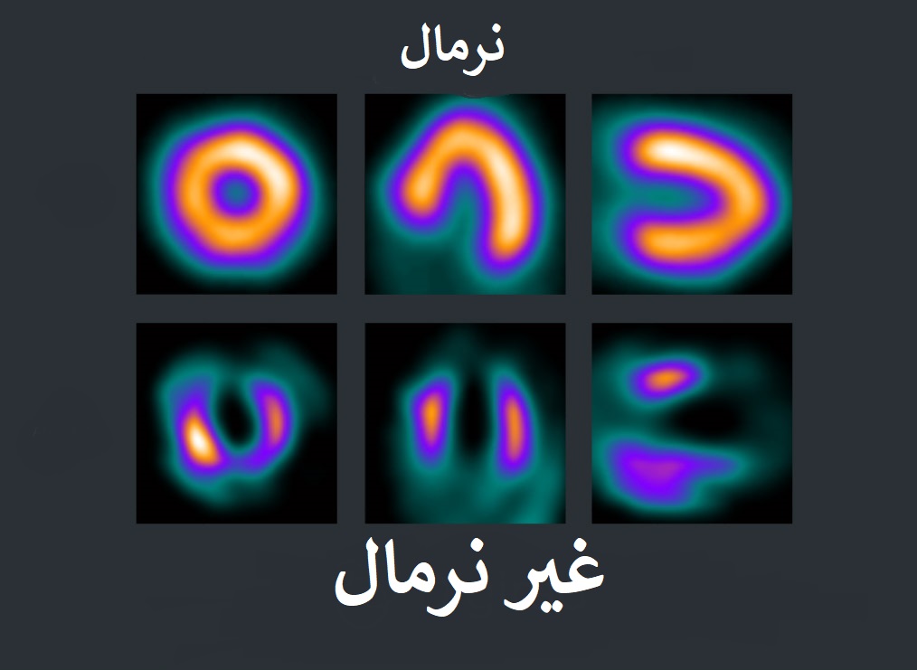